


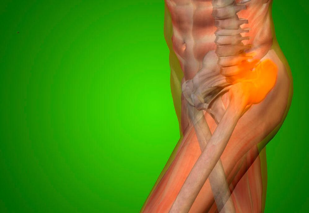
The hip joint experiences the greatest load on the body. They are created from the weight of walking, jumping, running, lifting and carrying heavy objects. Sufferers often feel pain in the hip joint. Orthopedists at specialized hospitals determine the cause using modern diagnostic equipment. Doctors determine the degree of joint damage, which allows them to make an accurate diagnosis and develop optimal treatment tactics.
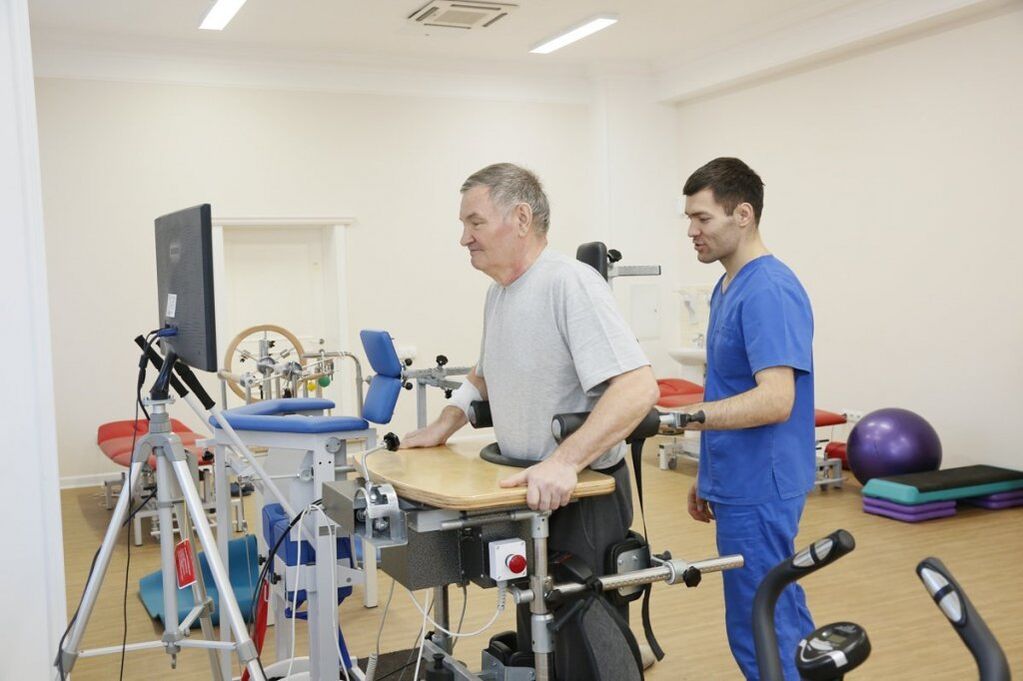
Professional doctors provide complex therapy for diseases that cause pain in the hip joint. The patient is individually selected effective drugs that affect the causes and mechanisms of pain development. Rehabilitation clinic specialists provide rehabilitation therapy using the latest physiotherapy, physical therapy and acupuncture procedures. The presence of special simulators allows you to reduce the load on the joints during exercise.
In the process of treating pain in the hip joint, doctors from various fields of medicine are involved: endocrinologists, rheumatologists, orthopedists, physiotherapists, chiropractors, acupuncturists. A multidisciplinary approach in the treatment of pain in the hip joint allows for rapid pain relief. Patients suffering from hip joint pathology often require external treatment.
Reason
Pain in the hip joint is caused by the following pathological processes:
- Tendinitis (tendon inflammation);
- Muscle rupture;
- iliotibial band syndrome;
- Other local changes in surrounding tissue;
- Systemic diseases (rheumatoid arthritis, polymyalgia).
Since the gluteus medius and minimus muscles play a major role in hip abduction, damage to them causes hip pain. The gluteus medius and minimus tendons attach to the greater trochanter. If an inflammatory process develops in them as a result of microtrauma due to excessive load, the patient will be disturbed by pain in the hip joint. The disorder may be caused by an infectious process (tuberculosis), sports or stereotypical professional stress, or the deposition of crystals.
Hip pain is a symptom of the following diseases:
- Osteoarthritis;
- Radicular syndrome;
- Rheumatoid arthritis;
- Coxita.
Pain in the hip joint can bother people who are overweight, have legs of different lengths, or have flat feet. Pain syndrome may occur after lower extremity amputation or hip replacement. With avascular necrosis of the head and fracture of the femoral neck, patients complain of acute pain in the hip joint. Pain syndrome often develops with dysplasia (disruption of the anatomical structure) of the hip joint. Acute pain in the hip joint, radiating to the leg, occurs in cases of pinched nerves due to spinal diseases, malignant bone tumors and age-related changes.
Inspection method
During the first consultation, the rheumatologist conducts a thorough examination of the patient:
- Collection of complaints, clarification of the nature of pain in the hip joint;
- Obtain information about the course of the disease, the onset of pain, the development of pain, household and professional factors that, in the patient's opinion, cause pain;
- External examination allows the doctor to determine visible deviations from the norm. To understand the nature of the pain and the extent of its spread, the doctor asks the patient to perform various movements of the lower limbs in the hip joint. The presence of hip joint pathology may indicate poor posture;
- Palpation (feeling). The doctor can find arthritic nodules and rheumatism, detect the exact location of pain when moving the legs, determine the humidity and temperature of the skin in the hip joint area.
Next, the doctor performs goniometry - an examination using a goniometer. This allows you to determine the range of joint mobility. Then the rheumatologist prescribes clinical and biological blood tests and a general urine test. Hospital laboratory technicians conduct research using high-quality reagents and modern equipment, so you can obtain accurate test results.
With inflammation of the hip joint, the number of leukocytes in the blood increases and the erythrocyte sedimentation rate increases. The inflammatory nature of the disease is indicated by an increase in the content of C-reactive protein in the blood serum.
Immunological blood test shows the presence of antinuclear antibodies in the blood in inflammatory rheumatic diseases. In arthritis sufferers, the concentration of uric acid in the blood serum increases sharply. The content of lysosomal enzymes (acid proteinase, acid phosphatase, cathepsin, deoxyribonuclease) in blood serum and changes in synovial fluid in patients with rheumatism, psoriatic polyarthritis, rheumatism and ankylosing spondylitis. In severe forms of hip joint pathology, significant deviations from the norm are observed in urine analysis.
Doctors at the clinic performed X-ray examinations on patients suffering from hip pain. This is demonstrated in the following cases:
- The presence of chronic or acute pain in the hip joint at rest and during movement;
- Difficulty in moving the lower limbs;
- The appearance of swelling and discoloration of the skin in the hip joint area.
Using computer tomography, doctors at the clinic evaluate the bones that play a role in the formation of the hip joint. On a computed tomogram, the radiologist finds changes in the structure of bone tissue, cartilage growth and osteophytes.
Using magnetic resonance imaging, the doctor evaluates the condition of the soft tissues surrounding the hip joint.
The radionucleotide research method allows recognizing pathology using radiopharmacological drugs.
Ultrasound examination of the hip joint is carried out to detect injuries, inflammatory diseases, rheumatism and rheumatoid arthritis. The attending physician individually selects the necessary research methods to determine the cause of pain in the hip joint in each case.
Differential diagnosis
Pain in the hip joint when walking is the main complaint of patients who consult a doctor. It can be located in the joint area or extend to the thighs, buttocks or knee joints. If pain occurs in the hip joint when moving, the patient is forced to use a cane. Often due to pain, there is limited mobility when moving the hip joint, especially when turning the leg outward and inward.
Pain in the hip joint, buttocks and groin is a symptom of aseptic necrosis of the femoral head. This disease is often associated with long-term use of hormonal drugs and alcohol abuse. As the deformity of the femoral head progresses, the mobility of the hip joint becomes limited. In the early stages of the pathological process, the range of motion may be normal.
Pain in the anterior hip joint and clicking sounds when moving the joint bother patients suffering from iliopectineal bursitis. Spreads to the thigh and is accompanied by paresthesia (tingling, burning, crawling) due to compression of the femoral nerve. The patient feels pain in the hip joint when flexing and extending the lower limb. Pain is also detected on deep palpation in the region of the femoral triangle (formation limited by the inguinal ligament, the outer edge of the long adductor muscle, the inner edge of the sartorius muscle).
Pain in the outer hip joint is a sign of iliotibial band syndrome. Accompanied by a clicking sound when moving, pain in the outer knee joint, which gets worse with movement.
Roth's myalgia is manifested by burning pain in the anterior outer part of the hip joint and thigh, which increases when walking and straightening the leg. Pain in the hip joint occurs with dysplasia. Over time, the patient develops a typical "duck" gait (he walks, walking from side to side).
Pain with coxarthrosis
Pain in the hip joint occurs with coxarthrosis, a disease characterized by degenerative processes in the bones that form the joint. More often this disease attacks elderly people. As we age, joint cartilage tissue loses its elasticity, becomes thin, and begins to break down. When the load on the joint increases, the thin cartilage tissue is destroyed. The articular surfaces of the bones rub against each other, resulting in aseptic inflammation.
Growths appear on the bones. They significantly limit joint movement. Deformation of the articular surfaces develops, resulting in severe pain. Treatment of this disease depends on the severity of joint damage. The doctor provides drug therapy. If it is ineffective, endoprosthetics or palliative treatment is performed.
After determining the cause of pain in the hip joint, the doctor begins to treat the disease that caused the pain syndrome. Cases of severe disease in which patients are bothered by pain in the hip joint are discussed at meetings of the expert council with the participation of professors, doctors and candidates of medical sciences, doctors of the highest category.
Treatment
An important condition for successful treatment of diseases that cause pain in the hip joint is to eliminate the factors that cause structural changes in the bones, cartilage and soft tissues in the joint area. For acute pain, hospital rheumatologists prescribe nonsteroidal anti-inflammatory drugs. The patient's well-being improves significantly with the use of local treatment methods - external application of gels and ointments, patches containing non-steroidal anti-inflammatory drugs. They reduce pain in the hip joint during inflammatory processes of soft tissues (tendinitis, bursitis, epicondylitis), after injuries.
If such therapy is not effective enough, the doctor injects glucocorticoids into the hip joint cavity. The joint space with deforming coxarthrosis is narrowed, it is difficult to enter it. Therefore, rheumatologists in specialized clinics carry out this procedure under X-ray control. In the presence of pain caused by inflammation of muscles and tendons, glucocorticoid hormones are injected into the periarticular tissue.
To improve the condition of the cartilage and reduce pain in the hip joint, chondroprotectors are used. The therapeutic course lasts several months. When spasms occur in the muscles involved in hip joint movement, muscle relaxants are prescribed to reduce skeletal muscle tone.
Drug therapy is complemented by physiotherapeutic procedures. They are of secondary importance in pain in the hip joint. The effectiveness of physiotherapeutic treatment methods is reduced due to their deep location. The severity of pain in the hip joint decreases after medium-wave ultraviolet irradiation.
In the presence of an inflammatory process, high-intensity centimeter wave therapy, infrared laser treatment and low-intensity UHF are performed. High intensity high frequency magnetic therapy, ozone therapy, shock wave therapy stimulate tissue recovery. The intensity of pain that occurs as a result of impaired blood circulation and nutrition of the hip joint is reduced under the influence of various types of electrotherapy (exposure to current) and ultrasound.
To reduce the load on the hip joint, rheumatologists recommend that patients use crutches in case of acute pain. After reducing the severity of the pain syndrome, the rehabilitation specialist performs therapeutic exercises. An individual set of exercises is developed for each patient to quickly restore lower extremity function. When the structures that play a role in the formation of the hip joint are destroyed, the pain can be so severe that the only method to eliminate it is to replace the joint with an endoprosthesis.
Nonsteroidal anti-inflammatory drugs are prescribed to relieve pain. Treatment depends on the disease affecting the hip joint. Patients are prescribed chondroprotectors for cartilage tissue damage. An orthopedic doctor prescribes effective medication, diet, and exercise to improve blood circulation in the joint, restore cartilage tissue, and maintain joint mobility. In severe cases, joint replacement with an endoprosthesis is necessary, which significantly improves the quality of life and eliminates pain.
Treatment with exercise therapy
The use of rehabilitation techniques in the treatment of the hip joint allows you to maintain its mobility, improve blood circulation in the joint, and accelerate the recovery of cartilage tissue. Specialists in the rehabilitation department select a set of physical therapy exercises taking into account the patient's joint disease. Rehabilitation classes are conducted daily under the supervision of an instructor. For rehabilitation therapy, special simulators are used, and physiotherapeutic procedures are prescribed in combination with physical education.
What diseases cause joint pain
Pain in the hip joint on the right or left side may be a manifestation of avascular necrosis. This disease develops mainly in men and attacks only one joint. Treatment consists of eliminating pain, restoring blood supply to the joint area, normal condition of the limb muscles, and maintaining joint function. Patients are prescribed painkillers and anti-inflammatory drugs, vitamins, physiotherapy procedures and therapeutic exercises. Patients are recommended to wear orthopedic shoes and use additional support when moving.
The cause of pain in the hip joint may be a purulent process. Primary purulent arthritis develops when there is a wound or injury and an infectious agent enters the joint cavity. Secondary purulent processes develop when sepsis or infectious agents enter the joint from surrounding tissues affected by the inflammatory process. To treat purulent arthritis, professional specialists carry out antibiotic therapy. If pus accumulates in the joint cavity, the hip joint is punctured, the contents are evacuated, and an antibacterial agent is injected into the joint cavity.
Bursitis is inflammation of the joint lining. To relieve pain, doctors prescribe injections of anti-inflammatory drugs and glucocorticoids. If purulent inflammation develops, the periarticular bursa cavity is cleaned. In severe cases, using endoscopic surgical techniques, the joint capsule, which has undergone permanent changes, is removed.
In osteoporosis, fractures of the femoral neck often occur. Patients are bothered by sharp and severe pain when moving in the hip joint, which radiates to the groin and inner thigh. His feet face outward. Bruising and swelling appear in the area of the hip joint. In this case, treatment is carried out by a professional orthopedist.
Traumatic hip dislocation is accompanied by pain in the hip joint. The hip is reduced under general anesthesia. Congenital hip dislocation is diagnosed soon after birth. It manifests itself as severe pain when the leg is extended and the knee is bent. Treatment is carried out using special orthopedic structures.
If you or someone you love experiences pain in the hip joint, you should not self-medicate. Seek professional medical help immediately. Patients with acute pain are usually hospitalized in a clinic for at least a week. If the pain is not intense, the patient may be offered an examination by a professional doctor for diseases of the hip joint and treatment at home with strict adherence to all the rules.